By Audrey Anderson
Hometown Weekly Reporter
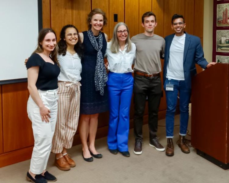
Collaboration of an Artist and a Scientist: Exploring Bones was presented on Monday evening, June 12, at the Needham Public Library. In this talk, Professor Sandra Shefelbine, of Northeastern University, and local artist Colleen Pearce discussed their recent combined scientific research and art creation project involving the study of bones.
Professor Shefelbine started the evening with an overview of the research that she and her graduate students have been conducting on factors that contribute to bone health and the body’s natural bone tissue functions.
Artist Colleen Pearce then explained how she observed the scientists’ presentations and research to gain insight into and inspiration from the structures they were examining. She then began her own explorations of what she saw in the lab through shape, structure, light, and color. Pearce’s resulting artwork was on display in the Friends of the Needham Public Library June 1 through June 15.
Ph.D. candidate Vineel Kondiboyina said his research “delves into the intriguing world of cartilage mechanics and its extraordinary ability to adapt and thrive.” In “exploring the intricate response of cartilage to mechanical forces during various stages of growth,” he has found that “cartilage, the smooth white tissue nestled between our joints, possesses an awe-inspiring mechanism of function regulation devoid of any blood supply or nervous tissue. Instead, it relies on the remarkable utilization of mechanical cues, such as the compression experienced during walking, to orchestrate its vital processes . . .
Calcium serves as a fundamental player in the cartilage's ability to adapt and maintain homeostasis . . . different speeds of loading, like walking versus running, influence calcium activity. To visualize these dynamic events, [Kondiboyina] employed fluorescent molecules tagged to calcium, enabling the cells to emit a vibrant green glow whenever calcium activity surges. This remarkable phenomenon, known as cell activation, bears immense significance, as it allows the cells to produce the proteins essential for tissue growth and preservation. . . . An inadequate load or an overwhelming burden can lead to debilitating conditions, such as osteoarthritis, a malady that currently lacks a cure. Understanding and manipulating these minute bursts of calcium become critical for maintaining the health and resilience of cartilage.”
Ph.D. candidate Quentin Meslier explained that “bones are able to adapt to load . . . To better understand how we form new bone in response to loading, we need to zoom into the bone cells and look at the molecules that they express. (“Molecules” is a general term that includes proteins, DNA, and everything in the cell[s] or made by the cells.) In the context of bone, cells use these molecules to control and regulate . . . bone formation.”
“I am developing methods to investigate the expression of these important molecules in . . . mouse bones. . . . It “is challenging because these molecules are tiny and they are located in . . . a dense mineralized tissue. . . . Our method allows us to make the mouse bones transparent . . . and we tag the important molecules of interest in the bone cells with fluorescent dyes. Then we place the bones in a microscope to image the fluorescent molecules . . . With these images we can have an idea of where the cells are located in the bone, and which cells express the important molecules. Using this method, the question that we are now asking is: does the expression of these molecules change in a leg that has been loaded, compared to a non-loaded leg? Is the expression of these molecules different in a young bone, compared to an older bone?”
Ph.D. Candidate Soha Ben Tahar added that “understanding how patterns emerge [in embryonic development] is crucial for unraveling the mysteries of embryogenesis. One key factor in this process is the role of morphogens, chemical signals that cells use to communicate with each other, which play a crucial role in determining cell fate and tissue specialization. These remarkable signals enable cells to organize themselves into complex patterns, even when they are a million times smaller than the final structure. Alan Turing proposed a model for how morphogens work, known as the reaction-diffusion system, which has been used to model patterning in a variety of biological applications. By studying how chemical signals interact and spread within the growing embryo, researchers hope to gain insights into how different tissues and organs take shape.”
Both Professor Shefelbine and artist Colleen Pearce observed how the scientific and artistic processes have much in common. Both processes utilize multiple experiments with different materials and approaches until they arrive at a result that is satisfactory.


